The superclass of fish belongs to the group of organisms that are in a state of pronounced biological progress. There are about 20 thousand species of fish living in the seas and fresh waters.
The emergence of fish was associated with the appearance of a number of aromorphoses:
- skulls are containers for the brain;
- jaws that provide active capture of prey;
- paired fins providing greater mobility;
- progressive development of the central nervous system.
Fish are animals adapted to narrow, rather monotonous living conditions - the aquatic environment, in which they differentiate into large number species.
Representatives of the superclass of fish
Let's look at the characteristic features of fish using the example of perch. In our country it lives everywhere (with the exception of Lake Balkhash and Far East) in rivers, lakes, reservoirs and flowing ponds. The body shape of the perch, like most other fish species, is streamlined, which allows it to better overcome water resistance.
There are three sections in the body of a fish:
- head;
- torso;
- tail.
The conventional boundary between the head and the body is the back of the gill covers, and between the body and the tail is the anus.
External structure
Coverings and body coloring. The entire body of the perch, with the exception of the head, is covered with bony scales. They are located in the right rows. The front edge of the scales is immersed in the skin, and the back edge overlaps the scales of the next row. Scales are a protective cover and do not interfere with the movements of the fish’s body.
The scales are covered on top with thin skin, the unicellular skin glands of which secrete abundant mucus. A layer of mucus reduces friction on the fish's body when swimming and protects it from pathogens of bacterial and fungal diseases.

The belly of the perch is lighter than the back. This is adaptive in nature, since the perch is less noticeable from below, against a light background of the water surface, and from above, against a dark background of the bottom. In lakes with a dark bottom there live perches with a dark color, and in reservoirs with a light sandy bottom - light ones, which is a consequence of the selection and survival of those most adapted to specific environmental conditions.
The greenish color of the perch and the vertical dark stripes on its body help it to be less noticeable in the thickets where it often lurks. The protective coloring allows them to better watch for prey and hide from enemies.
Skeleton and musculature of fish
Perch skeleton consists of the skull, spinal column and skeleton of the limbs (fins). The skull consists of two sections - the brain and the gill-maxillary:
- The cerebral part of the skull contains the brain, organs of smell, vision and hearing;
- The branchial-maxillary section consists of the bones of the upper and lower jaws, the hyoid and gill arches and the gill covers.

The spinal column is divided into trunk and caudal sections. It consists of 39-42 vertebrae. There are trunk and caudal vertebrae. Each vertebra has a body, on the dorsal side of which is adjacent the upper arch with the spinous process. Remnants of the notochord are preserved between adjacent vertebral bodies.
The spinal cord is located in the canal formed by the upper vertebral arches. In the trunk section, ribs extend from the vertebrae, and in the caudal section there are lower arches with lower processes. The skeleton of paired fins has belts and bony rays, while unpaired fins have only bony rays. There are two belts: shoulder and pelvic.
Musculature. The bulk of the muscles of a perch are located in the form of separate segments, which are connected to each other by fibrous layers. There are also specialized muscles (muscles of the pectoral and pelvic fins, gill cover, muscles that move the jaws, eyes). Forward movement carried out due to the work of the muscles of the caudal fin.
Internal organ systems
Digestive system. The alimentary canal begins with the oral opening, which leads into the oral cavity. On the jaws and other bones of the perch's oral cavity there are numerous undifferentiated teeth that serve only to capture and hold prey. Next comes the esophagus, stomach and intestine, ending with the anus. There is a liver. The pancreas is poorly developed.

swim bladder in perch, like in many other fish species, it is a hydrostatic apparatus. It is a thin-walled outgrowth of the intestine, which in the larvae of perch and some fish species (carp, roach) is connected to the intestine throughout life by a small tube.
The connection between the swim bladder and the intestine is lost in adult perch. The bladder is located above the intestine and is filled with gas, which includes oxygen, carbon dioxide and nitrogen. The walls of the bladder are penetrated by numerous blood vessels through which the blood releases or absorbs gas.

As the fish submerges or ascends, the amount of gas can increase or decrease, thereby regulating the fish's body density. Since the swim bladder has many blood vessels, which in some places form capillary accumulations, it can serve some fish burrowing in the mud for gas exchange.
They are represented by gills, consisting of gill arches, gill filaments and gill rakers. Gill rakers are a filtering apparatus that prevents swallowed prey from slipping out through the gill slits.

Gill filaments cover the gill arches like a fringe. The petals of living fish have a bright red color, due to the translucency of many small blood vessels. Gas exchange occurs in the gill filaments. The delicate gills cover the outside of the gill covers.
The excretory organs are the kidneys. Two ureters carry urine into the bladder, which opens to the outside behind the anus.
Circulatory system in fish represented by one circle of blood circulation. Only venous blood enters the heart. Fish have a two-chambered heart. It consists of the atrium and ventricle. In the gills, the blood is saturated with oxygen.

Nervous system in fish. The central nervous system of the perch consists of the brain and spinal cord. The brain is represented by five sections typical for vertebrates (anterior, intermediate, middle, cerebellum and medulla oblongata), but it still has a primitive structure. The forebrain is poorly developed. It serves as the highest olfactory center. The midbrain reaches its largest size. The cerebellum is well developed, which is associated with complex coordination of movements.

Pisces class- this is the largest group of modern vertebrates, which unites more than 25 thousand species. Fish are inhabitants aquatic environment, they breathe with gills and move with the help of fins. Fish are distributed in different parts of the planet: from high mountain reservoirs to ocean depths, from polar waters to equatorial ones. These animals inhabit the salty waters of the seas, found in brackish lagoons and estuaries large rivers. They live in freshwater rivers, streams, lakes and swamps.
External structure of fish
The main elements of the external body structure of a fish are: head, operculum, pectoral fin, pelvic fin, body, dorsal fins, lateral line, caudal fin, tail and anal fin, this can be seen in the figure below.
Internal structure of fish
|
Fish organ systems |
||
|
1. Skull (consists of the braincase, jaws, gill arches and gill covers) 2. Skeleton of the body (consists of vertebrae with arches and ribs) 3. Skeleton of fins (paired - pectoral and abdominal, unpaired - dorsal, anal, caudal) |
1. Brain protection, food capture, gill protection 2. Protection internal organs 3. Movement, maintaining balance |
|
|
Musculature |
Wide muscle bands divided into segments |
Movement |
|
Nervous system |
1. Brain (divisions - forebrain, middle, medulla oblongata, cerebellum) 2. Spinal cord (along the spine) |
1. Movement control, unconditioned and conditioned reflexes 2. Implementation of the simplest reflexes, conduction of nerve impulses 3. Perception and conduction of signals |
|
Sense organs |
3. Hearing organ 4. Touch and taste cells (on the body) 5. Lateral line |
2. Smell 4. Touch, taste 5. Feeling the direction and strength of the current, the depth of immersion |
|
Digestive system |
1. Digestive tract (mouth, pharynx, esophagus, stomach, intestines, anus) 2. Digestive glands (pancreas, liver) |
1. Capturing, chopping, moving food 2. secretion of juices that promote food digestion |
|
swim bladder |
Filled with a mixture of gases |
Adjusts immersion depth |
|
Respiratory system |
Gill filaments and gill arches |
Carry out gas exchange |
|
Circulatory system (closed) |
Heart (two-chambered) Arteries Capillaries |
Supplying all body cells with oxygen and nutrients, removing waste products |
|
Excretory system |
Kidneys (two), ureters, bladder |
Isolation of decomposition products |
|
Reproduction system |
Females have two ovaries and oviducts; In males: testes (two) and vas deferens |
Fish are aquatic vertebrates that breathe through gills. The limbs look like fins. The body of most fish is covered with scales. Body temperature depends on temperature surrounding water. The body shape is very diverse, but usually has a streamlined outline, which makes it easier for fish to move through water, a denser environment than air. The body is divided into head, trunk and tail. The movement of fish is carried out by bending the body and using fins. The fins are thin folds of skin supported by cartilaginous or bony rays.
Fig.1. Rudd (lat. Scardinius erythrophthalmus)
There are paired and unpaired fins. The first lie in the middle plane of the body - these are the caudal, dorsal (or dorsal) and anal fins. With the blows of the tail and caudal fin, the fish moves forward, and the dorsal and anal fins, like the keels of a boat, direct the movement of the body. Paired pectoral and pelvic fins serve as depth rudders and help the fish change directions of movement. The skin of fish is mucous, which reduces friction with the water. Most fish have skin covered with scales of different structures and shapes.
Nervous system divided into central and peripheral. The central nervous system is formed by the brain and spinal cord. The brain consists of 5 sections: the forebrain with the olfactory nerves extending from it, the diencephalon, from which the optic nerves go to the eyes, the midbrain, the cerebellum and the medulla oblongata. Each department performs specific functions in the nervous activity of the animal. The forebrain does not form hemispheres. The peripheral nervous system consists of a branched system of nerves that extend from the brain and spinal cord to all organs of the body.
Among the sense organs, fish have well-developed eyes, hearing aids, olfactory organs, and taste buds in the mouth. There is also a special sense organ - the lateral line. On the sides of the body there is a series of holes leading into a longitudinal channel lying in the skin. Its walls contain numerous nerve endings. Apparently, the lateral line organ perceives changes in pressure and movement of water.
The fish's mouth leads into the pharynx, in the side walls of which there is a series of gill slits. In most fish, the slits are separated by bony or cartilaginous gill arches, on which red thin gill filaments sit on the outside, and whitish gill rakers on the inside. The fish swallows water, which washes the petals of the gills and comes out. In this case, the oxygen contained in the water penetrates into the blood. The gill rakers form a filtering apparatus that prevents food swallowed by the fish from exiting through the gill slits. Food swallowed by fish passes through the esophagus into the stomach, where it is exposed to gastric juice and begins to be digested. Further digestion of food occurs in the intestines, digested food is absorbed by the intestinal walls, undigested food remains are thrown out through the anus.
Most fish have a swim bladder filled with a mixture of gases in their body cavity. By contracting and expanding, it changes the volume, and therefore the density of the animal, which is always equal to or very close to the density of the environment.
Circulatory system. There is only 1 circle of blood circulation. From the heart, which is made up of 2 sections - the atrium and the ventricle, venous blood flows to the gills, where it is enriched with oxygen and freed from carbon dioxide. From the gills, arterial blood spreads through the arteries throughout the body. Venous blood flows through the veins to the atrium.
Excretory organs. The excretory organs of fish are 2 kidneys, located under the spine in the body cavity. The urine they secrete flows down two ureters into the bladder or directly out.
Skeleton of fish. The axial skeleton can be represented by a notochord or a vertebral column. In cyclostomes, sturgeons and lungfishes, the notochord is retained throughout life. In all other fish, the notochord is present in the early stages of development, and in adults it is replaced by a spine consisting of vertebrae. The cranium is connected to the spine motionlessly. Fish have no neck. This is caused by the specifics of the lifestyle and habitat - the need to cut the water with your head. In the process of evolution, the skeleton became more complex and ossified. In cyclostomes, the notochord stretches from the back of the skull to the tail in the form of a solid, non-segmented cord consisting of cartilaginous and connective tissue elements (dorsal string), to which the cartilaginous vertebral arches are tightly adjacent on top. The notochord of sturgeons is also not yet differentiated. In elasmobranch (shark) fishes, the cartilaginous shell of the notochord forms amphicoelous (biconcave) vertebrae. Bony fish have an already ossified spine.

Rice. 2. Skeleton of a bony fish (perch) (according to Baklashova, 1980)
1 – skull bones, 2 – main elements of the dorsal fin, 3, 4 – rays of the dorsal fin, 5 – last vertebrae holding the caudal fin, 6 – caudal vertebrae; 7 – main elements of the anal fin, 8 – trunk vertebrae, 9 – ribs with appendages, 10 – bones and rays of the ventral fin, 11 – bones and rays of the pectoral fin, 12 – operculum, 13 – upper and lower jaws
It contains the trunk and caudal sections. The trunk section is divided into typical vertebrae - amphicoelous, which distinguish the body, the upper arch with the upper (neural) spinous processes (protecting the spinal cord) and the large lower arches with the lower processes. In the trunk region, the ribs are attached to the spine (to the transverse processes or to the vertebral body). In the caudal region, the transverse processes, closing, form the lower (hemal) arch, which ends in the lower spinous process. The hemal canal contains the caudal artery and vein. The last caudal vertebra is flattened and serves to attach the rays of the caudal fin; it often changes its usual shape: it lengthens and bends upward, forming a urostyle.
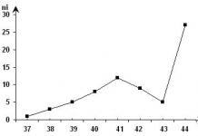
The number of vertebrae is determined by a number of internal and external factors and serves as a systematic feature of the fish. For example, northern herring has 57, river eel - 114, catfish - 72, sunfish - 17, pike perch - 44. Within a species, the dependence of the number of vertebrae (and rays in the pectoral and anal fins) on temperature is known: an increase in temperature during the period embryogenesis causes a decrease in their number. In addition to the ribs, the supporting function in bony fish is performed by thin “muscular” – intermuscular, or “trunk”, bones that penetrate the muscles. These bones are formed by ossified tendons. Most of them are found in carp fish.
Reproduction. Almost all fish are dioecious. In females, the body cavity contains an ovary, where eggs develop, and in males, there are testes, which produce a huge number of sperm. The vast majority of fish lay eggs, but there are some that will give birth to live young.
The development of the genitourinary system in the evolution of fish led to the separation of the reproductive ducts from the excretory ducts. Cyclostomes do not have special reproductive ducts. From the ruptured gonad, the sexual products fall into the body cavity, from it - through the genital pores - into the urogenital sinus, and then through the urogenital opening they are discharged out. In cartilaginous fish, the reproductive system is connected to the excretory system. In females of most species, eggs are released from the ovaries through the Müllerian canals, which act as oviducts and open into the cloaca; The Wolffian canal is the ureter. In male wolfs, the canal serves as the vas deferens and also opens into the cloaca through the urogenital papilla. In bony fishes, the Wolffian canals serve as ureters, the Müllerian canals are reduced in most species, and reproductive products are excreted through independent genital ducts that open into the genitourinary or genital opening. In females (most species), mature eggs are released from the ovary through a short duct formed by the ovarian membrane.
In males, the testicular tubules connect to the vas deferens (not connected to the kidney), which opens outward through the genitourinary or genital opening. Sex glands, gonads - testes in males and ovaries or ovaries in females - ribbon-like or sac-like formations hanging on the folds of the peritoneum - mesentery - in the body cavity, above the intestines, under the swim bladder. The structure of the gonads, which are similar at the core, has some peculiarities in different groups of fish. In cyclostomes, the gonad is unpaired; in true fish, the gonads are mostly paired. Variations in gonad shape various types are mainly expressed in the partial or complete fusion of paired glands into one unpaired one (female cod, perch, eelpout, male gerbil) or in a clearly expressed asymmetry of development: often the gonads are different in volume and weight (capelin, silver carp, etc.), up to until one of them completely disappears. From the inner side of the walls of the ovary, transverse egg-bearing plates extend into its slit-like cavity, on which germ cells develop. The basis of the plates is made up of connective tissue cords with numerous branches. Highly branched blood vessels run along the cords.
Mature reproductive cells fall from the egg-laying plates into the ovarian cavity, which can be located in the center (for example, perch) or on the side (for example, cyprinids). The ovary directly merges with the oviduct, which carries the eggs out. In some forms (salmon, smelt, eels), the ovaries are not closed and mature eggs fall into the body cavity, and from there through special ducts they are removed from the body. The testes of most fish are paired sac-like structures. Mature germ cells are excreted through the excretory ducts - vas deferens - into external environment through a special genital opening (in male salmon, herring, pike and some others) or through the urogenital opening located behind the anus (in males of most bony fish).
The class of fish is divided into a number of systematic groups:
cartilaginous fish. These include sharks and rays - marine fish with a cartilaginous skeleton. The body is covered with special scales with sharp teeth protruding outward. Caudal fin with large upper and small lobes. There is no operculum; the gill slits open on both sides of the body with separate openings. Sharks' jaws are armed with sharp teeth. Stingrays live at the bottom of the seas.
osteochondral fish. These include sturgeon, beluga, stellate sturgeon, sterlet and other sturgeon fish. The notochord lasts throughout life. The internal skeleton is cartilaginous, but the outside of the head is covered with flat bones. There is an operculum.
teleosts make up the main group of modern fish. They differ in that in adult individuals the notochord is preserved in separate sections between the vertebrae, the skeleton is mainly formed by numerous bones, and the scales look like thin plates overlapping each other.
Bony fish include carp, crucian carp, roach, bream, perch, pike, ruffe, catfish, pike perch, etc.
A fish consists of a food tract and glands. Fish are divided into omnivores, detritivores, predators and herbivores. Herbivores eat microalgae, aquatic flowering plants, and plankton. Some marine species feed on macroalgae. Predators use crustaceans, mollusks, worms and small fish, parasites and dead parts of other fish for food resources. Some species change their diet throughout their lives, switching from plankton to fish and invertebrates. The types, work and importance of the digestive system in fish will be discussed in this article.
General information
Most fish have mouths equipped with many conical teeth designed to capture and hold food. It is not delimited by the pharynx, which leads into the short esophagus. The stomach has a variety of shapes and sizes. Its ability to stretch in deep-sea predatory fish helps them stock up on food.
Partial processing of food in the stomach occurs under the influence of juice produced by small glands of the mucous membrane. Food is completely processed and absorbed in the small intestine. To increase the surface of the digestion, there are blind processes in its upper part. The secretion produced by the pancreas through the ducts flows into the initial section of the intestine. Bile also goes there. Undigested food debris accumulates in the hindgut and is expelled through the anus.
Digestive tract
In fish, it consists of five sections: the oral cavity, pharynx, esophagus and stomach, which serves for partial digestion of food. In addition, there is an intestine in which the final absorption of food and nutrients occurs and waste is removed through the anus. The quality of nutrition is reflected in the digestive system of fish, and it can vary significantly between different breeds. Largemouth species have a mouth in the form of a special suction funnel, equipped with teeth inside. Fish stick to their prey, drilling into the body with a sharp tongue. They secrete a substance into the wound that partially dissolves the protein. Partially processed food enters the gastrointestinal tract.
The mouth, part of the digestive system, in predator fish is a grasping tool with sharp teeth, which are often arranged in several rows and firmly hold the prey.

Teeth do not have roots and do not last long, and then new ones grow. They can be located not only on the jaw, but also in other places in the oral cavity, including on the tongue. Predators have sharp teeth that are curved back; they are even in the throat. And many peaceful breeds have no teeth at all.
Pharynx
From the mouth, food enters the next section of the fish’s digestive system - the pharynx. It contains gill slits that open outward. There are stamens on the gills.

They help predators hold prey and protect the gills from damage. Other fish filter water through the latter and retain food. In addition, there are glands in the pharynx that produce mucus, which facilitates swallowing.
Esophagus and stomach
The pharynx passes into the esophagus, which is a small organ of the fish’s digestive system, which passes into the stomach, which helps move food. In pufferfish, the esophagus can swell, filling with air. In sharks, rays and salmon it consists of two sections, and in perches it is a blind process. Some aquatic inhabitants do not have a stomach at all, and the esophagus is replaced by intestines.
Intestines
The esophagus passes into the intestine, which begins with the small intestine. Two ducts flow into it from:
- the liver, which delivers bile;
- pancreas with catalysts (enzymes).
These components contribute to the breakdown of proteins into amino acids, fats into fatty acids and glycerol, and polysaccharides into sugars. After digestion, nutrients are absorbed into the blood through the folded walls of the stomach, equipped with outgrowths and penetrated by lymphatic vessels and capillaries. A feature of the digestive system of fish is the length of the intestines. It is directly dependent on the calorie content of food. In predators it is short, but in fish that feed on plankton it is long. The intestine of the silver carp is 16 times longer than its own size. In all species of fish, it ends with the anus, located between the genital and urinary passages.
Liver and pancreas
Considering the structure of the digestive system of fish, it should be said that it includes the liver and pancreas. The enzymes they produce pass through the ducts into the small intestine. Cartilaginous fish species have a three-lobed liver, while bony fish have one to three. It makes up up to 20% of the total weight of the fish. Bile produced by the liver stimulates the intestines, breaking down fats. This organ neutralizes toxic substances, synthesizes carbohydrates and proteins, accumulates vitamins and glycogen. All this is necessary for adequate functioning of the body. The pancreas synthesizes enzyme substances that affect food processing. In addition, it produces insulin, which regulates the concentration of glucose in the blood. In some fish (for example, cyprinids), pancreatic tissue is contained in the liver. In fish that feed on plant foods, the process of processing nutrients involves microorganisms located in the intestines and secreting enzymes.
Digestive system of cartilaginous fish
Cartilaginous fish are mainly inhabitants of salt waters, but some species also exist well in fresh water. This species is a predator and feeds on small relatives and bottom-dwelling crayfish, crabs, mollusks, and sometimes jellyfish. Due to the special shape of the body, cartilaginous fish move quickly and have jaws with very sharp teeth; there is no real tongue.

Their skeleton consists entirely of cartilage, there are no gill covers and there is no swim bladder. The digestive system of cartilaginous fish consists of an oral cavity, a pharynx with gill slits, a short esophagus with muscular walls, and a stomach. The breakdown and absorption of food occurs in the small intestine, which has a folded inner surface and a short length that ends at the anus. Cartilaginous fish retain a cloaca. The functions of the digestive system of fish are performed by the liver, gallbladder and pancreas. They produce enzymes that travel through ducts into the upper part of the small intestine and help break down nutrients into amino acids, vitamins and fatty acids. It should be noted that the pancreas in cartilaginous fish is an independent organ and is located separately, and the large liver consists of three lobes and accounts for up to 20% of body weight.
Features of digestion of bony fish
The digestive system of bony fish has the same sections as those of cartilaginous fish. The location of the mouth opening depends on the type of food. The oral cavity contains many teeth. All of them are of the same type, inclined towards the pharynx and are adapted only for capturing and holding food. The pharynx with gill slits takes an active part in the feeding process. In some species of fish, in the pharynx on the gill arch there are powerful and large pharyngeal teeth for grinding food. The short esophagus passes into the stomach, which is not present in all fish species.

After the stomach, food enters the small intestine. At its very beginning there are the ducts of the liver and pancreas. This is where food is broken down and absorbed. In bony fishes the small intestine is much longer than in cartilaginous fishes. It forms loops, which increases the suction surface. Many types of fish, when the stomach passes into the small intestine, have appendages for the breakdown of protein and the absorption of amino acids. Bony fish have a gall bladder and a multi-lobed liver, which can consist of 1-3 lobes. The structures of the pancreas, unlike cartilaginous fish, are located in the liver tissue. The small intestine smoothly passes into the large intestine and ends with the anus.
Features of the digestive system of sturgeon fish
Sturgeons have a digestive tract that, in its structure, occupies an intermediate place between cartilaginous and bony fish. All sturgeons have a retractable jaw apparatus, representing an oral funnel. Its ability to extend far outward allows it to collect food from the bottom of the reservoir. Only larvae have teeth. In adults, they are replaced by horny ridges. In the digestive system of sturgeon fish, there are two adaptations that increase the absorption surface area:
- like in bones, the intestine creates several loops;
- like cartilaginous ones, the midguts retain a spiral fold.

In sturgeons, on the intestinal walls, fused appendages form the pyloric gland, which opens into the intestine. Instead of a cloaca, there are two openings: the genitourinary and the anal. The digestive tract has the same sections as in vertebrates. Liver, which has irregular shape, occupies the entire front part of the body and consists of three lobes. The shape of the gallbladder is oval. Its ducts empty into the duodenum. The pancreas is an independent organ.
Classification of fish by type of digestive system
The digestive tract of fish is much simpler in structure than that of higher vertebrates. But the wide variety of fish species and the structural features of their gastrointestinal tract are still not fully understood. To facilitate work in this direction, all types of fish according to the type of digestive system were divided into:
- salmon - stomach with thin walls, pyloric appendages from 80 to 400;
- perch - the walls of the pharynx are thick, the stomach is cylindrical, 3 pyloric appendages;
- pike - esophagus with thick walls, elongated stomach, liver elongated according to the geometry of the body;
- carp - there is no stomach, the digestive tract is a thin tube consisting of several loops, there is an extension in the upper intestine;
- acne - the esophagus is narrow and muscular, surrounded by the liver, it has several layers: mucous, submucosal, muscular, serous, ciliated.
Conclusion
At first glance, digestion in fish is simple. In fact, when studying this issue in detail, it turns out that there are many adaptations that help them obtain and absorb food. Individual groups of fish evolved under very different conditions.

Some of them ended up in fresh water, and the other in sea water. There are species of fish that can exist in salty and freshwater environments. Climatic conditions also differ greatly. As a result, different regions have completely different food supplies, to which the fish were forced to adapt. All this has led to a diversity of species and their digestive systems.
The internal structure of fish is considered using the example of river perch.
Musculoskeletal system. The basis of the internal skeleton of a fish (Fig. 117) is the spine and skull.
Rice. 117. Skeleton of a bony fish: A - general view: 1 - jaws; 2 - skull; 3 - gill cover; 4 - shoulder girdle; 5 - skeleton of the pectoral fin; 6 - skeleton of the ventral fin; 7 - ribs; 8 - fin rays; 9 - vertebrae; B - trunk vertebra; B - caudal vertebra: 1 - spinous process; 2 - upper arc; 3 - lateral process; 4 - lower arc
The spine consists of several dozen vertebrae, similar to each other. Each vertebra has a thickened part - the vertebral body, as well as the upper and lower arches. The upper arches together form the canal in which the spinal cord lies (Fig. 117, B). The arches protect him from injury. Long spinous processes protrude upward from the arches. In the trunk region, the lower arches (lateral processes) are open. The ribs are adjacent to the lateral processes of the vertebrae - they cover the internal organs and serve as a support for the trunk muscles. In the caudal region, the lower arches of the vertebrae form a canal through which blood vessels pass.
A small braincase, or skull, is visible in the skeleton of the head. The bones of the skull protect the brain. The main part of the head skeleton consists of the upper and mandibles, bones of the eye sockets and gill apparatus.
Large gill covers are clearly visible in the gill apparatus. If you lift them, you can see the gill arches - they are paired: left and right. Gills are located on the gill arches. There are few muscles in the head; they are located in the area of the gill covers, jaws and on the back of the head.
There are skeletons of unpaired and paired fins. The skeleton of unpaired fins consists of many elongated bones embedded in the thickness of the muscles. The skeleton of the paired fin consists of the skeleton of the belt and the skeleton of the free limb. The skeleton of the pectoral girdle is attached to the skeleton of the head. The skeleton of the free limb (the fin itself) includes many small and elongated bones. The abdominal girdle is formed by one bone. The skeleton of the free pelvic fin consists of many long bones.
Thus, the skeleton provides support for the body and organs of movement and protects the most important organs.
The main muscles are located evenly in the dorsal part of the fish's body; The muscles that move the tail are especially well developed.
swim bladder- a special organ characteristic only of bony fish. It is located in the body cavity under the spine. During embryonic development, it appears as a dorsal outgrowth of the intestinal tube (Fig. 118). The swim bladder prevents the fish from drowning under its own weight. It consists of one or two chambers, filled with a mixture of gases similar in composition to air. In so-called open-bladder fish, the volume of gases in the swim bladder can change when they are released and absorbed through the blood vessels of the bladder walls or when air is swallowed. This changes the volume of the fish's body and its specific mass. Thanks to the swim bladder, the body mass of the fish comes into balance with the buoyant force acting on the fish at a certain depth.

Rice. 118. Internal structure of bony fish (female perch): 1 - mouth; 2 - gills; 3 - heart; 4 - liver; - gallbladder; 6 - stomach; 7 - swim bladder; 8 - intestines; 9 - brain; 10 - spine; 11 - spinal cord; 12 - muscles; 13 - kidney; 14 - spleen; 15 - ovary; 16 - anus; 17 - genital opening; 18 - urinary opening; 19 - bladder
Digestive system begins with a large mouth located at the end of the head and armed with jaws. There is an extensive oral cavity. There are teeth. For oral cavity the pharyngeal cavity is located. It shows gill slits separated by interbranchial septa. They contain gills - the respiratory organs. Next comes the esophagus and a voluminous stomach. From the stomach, food enters the intestine. In the stomach and intestines, food is digested under the influence of digestive juices: in the stomach there is gastric juice, in the intestine - juices secreted by the glands of the intestinal walls and pancreas, as well as bile from the gallbladder and liver. In the intestines, digested food and water are absorbed into the blood. Undigested residues are thrown out through the anus.
Respiratory system located in the pharynx (Fig. 119, B, C). The skeletal support of the gill apparatus is provided by four pairs of vertical gill arches, to which the gill plates are attached. They are divided into fringed gill filaments. Thin-walled blood vessels branching into capillaries run inside them. Gas exchange occurs through the walls of capillaries: absorption of oxygen from water and release of carbon dioxide. Water moves between the gill filaments due to the contraction of the pharyngeal muscles and the movement of the gill covers. On the side of the pharynx, bony gill arches bear gill rakers. They protect the soft, delicate gills from becoming clogged with food particles.

Rice. 119. Circulatory and respiratory systems of bony fish: A - diagram of the circulatory system: 1 - heart; 2 - abdominal aorta; 3 - afferent gill arteries: 4 - efferent gill arteries; 5 - carotid artery (carries blood to the head); 6 - dorsal aorta; 7 - cardinal veins (carry blood to the heart); 8 - abdominal vein; 9 - capillary network of internal organs: B - gill arch: 1 - gill rakers; 2 - gill filaments; 3 - gill plate; B - breathing pattern: 1 - direction of water flow; 2 - gills; 3 - gill covers
Circulatory system closed fish (Fig. 119, A). Blood continuously flows through the vessels due to the contraction of the two-chambered heart, consisting of an atrium and a ventricle. Venous blood containing carbon dioxide passes through the heart. When the ventricle contracts, it directs blood forward into a large vessel - the abdominal aorta. In the region of the gills, it splits into four pairs of afferent gill arteries. They branch capillaries forward in the gill filaments. Here the blood is freed from carbon dioxide, enriched with oxygen (becomes arterial) and is sent through the efferent branchial arteries to the dorsal aorta. This second large vessel carries arterial blood to all organs of the body and to the head. In organs and tissues, the blood releases oxygen, is saturated with carbon dioxide (becomes venous) and enters the heart through the veins.
Nervous system. The central nervous system (CNS) consists of the brain and spinal cord (Fig. 120, A). The brain has five sections: the forebrain, diencephalon, midbrain, cerebellum and medulla oblongata (Fig. 120, B).

Rice. 120. Nervous system of bony fish: A - general scheme: 1 - cranial nerves; 2 - brain; 3 - spinal cord; 4 - spinal nerves; B - diagram of the brain: 1 - forebrain; 2 - diencephalon; 3 - midbrain; 4 - cerebellum; 5 - medulla oblongata
The medulla oblongata smoothly passes into the spinal cord. The peripheral nervous system is represented by nerves connecting the central nervous system to organs. The cranial nerves arise from the brain. They ensure the functioning of the senses and some internal organs. The spinal nerves arise from the spinal cord. They regulate the coordinated functioning of the muscles of the body, organs of movement, and internal organs. The nervous system coordinates the activities of the entire organism and the animals’ adequate reactions to environmental influences.
Excretory organs represented by the kidneys located along the spine, the ureters and the bladder (see Fig. 118). Through these organs, excess salts, water and waste products harmful to the body are removed from the fish’s body.
Urine flows through the ureters into the bladder and is expelled from it.
Laboratory work No. 7
Subject. Internal structure of fish.
Target. To study the features of the internal structure of fish and its complexity in comparison with skullless animals.
Equipment: tweezers, bath, ready-made wet fish preparation (or opened fresh fish).
Work progress
- Consider the location of the internal organs in the body of the fish.
- Find and examine the gills. Determine their location. Determine which organ system they belong to. How do fish breathe?
- Find the stomach, intestines, liver.
- Find the heart on the wet preparation. Determine its location in the body cavity. What organs belong to the circulatory system? Why is this circulatory system called closed?
- Determine whether you are considering a female or a male. Establish the location of the testes (ovaries) in the body cavity.
- Determine the location of the kidneys in the body cavity. Indicate which organ system the considered organs belong to. How are harmful waste products removed from the fish’s body?
- Draw a conclusion.
Compared to lancelets, fish are more highly organized animals. Their notochord is replaced by a spine; gills have a complex structure; the heart is muscular, two-chambered; The excretory organs are the kidneys, ureters and bladder. The central nervous system (neural tube) is divided into the brain (five sections) and the spinal cord.
Exercises based on the material covered
- Name the main parts of the fish skeleton. What function do they perform?
- What organs make up the musculoskeletal, respiratory, circulatory, central nervous system fish?
- List characteristic features internal structure of fish.
- Explain the importance of the swim bladder in the life of bony fish.


